

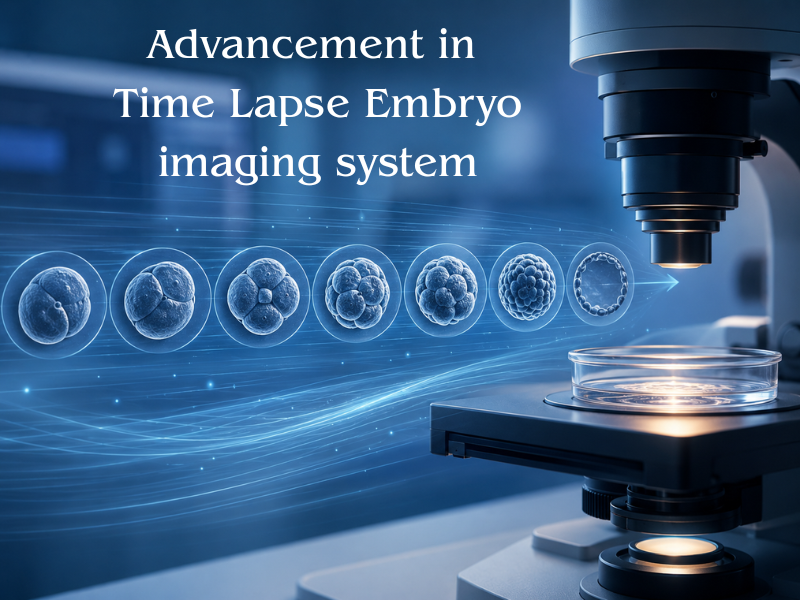
Time-lapse embryo imaging has revolutionized the way embryologists observe and evaluate embryo development. By capturing images at frequent intervals, these systems create a continuous video of embryonic growth, providing unprecedented insights into morphokinetics.
The Advantage of Uninterrupted Culture
Traditional assessment required removing embryos from the incubator, exposing them to fluctuations in temperature, pH, and light. Time-lapse incubators eliminate this need. Embryos remain in a stable, optimized environment throughout their development, reducing stress and potentially improving viability.
Morphokinetic Markers
The detailed developmental timelines generated by time-lapse systems allow embryologists to identify specific morphokinetic markers associated with higher implantation potential. For instance, the exact timing of cell divisions and the duration of specific developmental stages provide valuable predictive data.
With integrated software and increasingly sophisticated algorithms, time-lapse imaging is becoming an indispensable tool in the modern IVF laboratory.